Wednesday, October 2, 2013

Day-Bailes tapered locking suction tube system: Review.

Day-Bailes tapered locking suction tube system - recommended.

Pros: I particularly like this suction system which we have been using for around two years. It balances well in the hand, is malleable and is good for doing precise work.
The scrub nurse can switch the cannulas pretty fast and there is a wide range of sizes.

Cons: One needs to get used to the extra stuff that sits on the web space. This may not be so useful for extra dural work.

Downtime/Repairs: None so far
Institute where it was used: Jipmer, Pondicherry.

Price: Around INR 30 k.


Vendor's website: http://www.mizuho.com/day-bailes-locking-suction-tubes


Specifications( if you need to put in a tender)

Balanced handpiece (Flat teardrop suction handle with safety coupling and  suction tube handle) which can be kept between thumb and forefinger space for a attaching a range of suction cannulas.

Balanced handpiece (Flat teardrop suction handle with safety coupling cannulas) should be of the following sizes (3 to 12 french, each in following lengths - Short: 7.6 cm Medium: 9.5 cm Long: 11.4 cm Extra long: 13.3 cm. Also multihole tube type each 5, 7 and 9 french (all suction cannulas [43 numbers] should preferably come in its own case). The suction cannulas have to be pre-bent 30 degrees, malleable and rotatable and should have the ability to change tubes without discarding hand piece.

Recommendation levels possible: Highly recommeded, Recommended, Not recommended.
Conflict of interests/disclosures: none.

Why Microneurosurgery?


This picture was photographed to scale. The size of the human head at the level of the posterior fossa is compared to the human hand. Ergo, microneurosurgery!

The other day one of the anesthesia residents asked why neurosurgeons use an operating microscope. This picture shows the comparative dimensions of the posterior fossa with respect to the human hand. It's now easy to understand that tight spaces require magnification to get rid of "small tumors" especially when such tumors are enveloped by eight cranial nerves and four critical vessels.

Thursday, October 27, 2011

World Stroke Day 2011

29 th October 2011 is World Stroke Day

Point your smart phone with a qrcode reader application on to this cube to activate link. Feel free to download this design  for stroke day campaigns at your hospital.

The World Stroke Organization (WSO) is calling for urgent action to address the silent stroke epidemic by launching the “1 in 6” campaign on World Stroke Day, 29 October 2010.

The “1 in 6” campaign celebrates the fact that not only can stroke be prevented, but that stroke survivors can fully recover and regain their quality of life with the appropriate long-term care and support. The two-year campaign aims to reduce the burden of stroke by acting on six easy challenges:

1. Know your personal risk factors: high blood pressure, diabetes, and high blood cholesterol.

2. Be physically active and exercise regularly.

3. Avoid obesity by keeping to a healthy diet.

4. Limit alcohol consumption.

5. Avoid cigarette smoke. If you smoke, seek help to stop now.

6. Learn to recognize the warning signs of a stroke and how to take action.

You can read further at http://www.worldstrokecampaign.org/2011/Pages/Home.aspx

The  qr code  was generated using http://qrcode.kaywa.com and then transforming the code using GIMP to create this cube design. Isn't it interesting that the qr code has a large error tolerance (redundancy of information or function) that it allows a custom design to be placed in it without loss of readability... much like the human brain!

Sunday, August 28, 2011

Mother ship is calling you home! Will you be there on 16 th December 2011?

NIMHANS is organising an alumni meet on Dec 16 th 2011.
You can click on picture above or download your invitation here.

Tuesday, July 12, 2011

How many millihelen is your craniotomy?

Beauty, they say, lies in the eyes of the beholder. If one wants to be technically accurate, aesthetic appreciation centre of the human brain is perhaps the medial orbito-frontal cortex [neuro­bi­ol­o­gist Semir Zeki's fMRI study]. Human brain appreciates beauty looking primarily for symmetry and congruence when it compares a given pattern with those in its memory that once evoked a pleasurable sensation.

So if one Helen is the unit of beauty that could launch a thousand ships, how many millihelen is your craniotomy?

So, what do we do routinely to preserve aesthetics in neurosurgery?

Shaveless craniotomy, sinusoidal stealth incision, bone flap fixation implants, burrhole buttons, minimal access or natural foramen access (endoscopic endonasal), or may be, avoid a scalp incision altogether as in radiosurgery/endovascular surgery…to name a few

How important do you think aesthetics is in neurosurgery (assuming equivalent care inside the brain too) and could you suggest a few tips which one could use in routine practice? Or do you think it is not that much of a concern as in Westerners? I myself had many patients preferring to have a ‘full head shave’!

Cheers,
Gopalakrishnan.

Responses copied from yahoo group site:

Re: [neurosurgery_nimhans] How many millihelen is your craniotomy?

Very interesting topic of discussion I must say. I agree about the cosmetic benefits of non-shaved craniotomy. Wound care does however become a little more frustrating...and people often complain of increased wound infection rates. At TMH I did a small prospective study looking at infection rates following non shaved surgeries. we had about 75 tumor craniotomies. We also looked at scalp swabs before and after scrubbing in these patients....and I devised a VAS based 4 point questionairre for assessing cosmetic outcome also...The results are still being anlysed and maybe I can update you all in a short while. On preliminary look we did not find a higher infection rate.
If I may ask, what are the wound infection rates at your centres....though I don't have exact figures my experience shows it to hover around the 5-10% mark. A lot of them are related to wound leaks and I have a feeling (though no objective evidence) that by fixing the bone flap the leaks decrease and so also the infections.
Also, what antibiotic policy do you all follow (which antibiotic and for how long). These may sound very mundane issues, but I think they are as much crucial to the overall outcome .

Ali

Sent: Wednesday, July 13, 2011 1:25 PM
Subject: Re: [neurosurgery_nimhans] How many millihelen is your craniotomy?
 
Hi Gopal,
this is a good topic u asked for.
We started practising preserving hair for all kinds of craniotomy since 4 yrs and the response from the patients is very good especially from the xx chromosomes.
some of the patients went back to working early because of good aesthetic look because u have taken care of it.
regarding burrhlole buttons -- the cost factor adding to overall budget makes us use it less as compared to preserving hair. some patients do complain of deppression at burrhole sites especially chronic SDH.
bone fixation --- frontal region -yes good for cosmesis and other areas depending on the budget.

overall if u take care of cosmesis for the people, its an additional bonus.
  Dr.Praveen Ganigi
MBBS MCh Neurosurgery
Consultant Neurosurgeon & Spine Surgeon
Manipal Hospital,
HAL Airport road
Bangalore-560017                                                                   

Sunday, April 24, 2011

Juha Hernesniemi, Ginde oration, Bombay hospital, 2011

Battling thousands of aneurysms at minus 22 degrees. What does that turn you into?

A steady handed super-vascular surgeon who clips them in the blink of an eye or in short - Juha Hernesniemi.

A focused and fast approach to common aneurysms is  the message he tried to convey. These are the few points I could gather

A standardized limited lateral supraorbital approach using a hemi-arc of the classical pterional craniotomy incision takes care of most of the anterior circulation aneurysm. [estimated time for craniotomy: 15- 20 minutes]

Operative site preparation is with a few  gentle swabbing of povidone after a limited shave. No time wasted there compared to hyper -enthusiastic scrubbing with multiple agents over and over. After all it is just the contact time of five minutes that kills pathogenic bacteria rather than one's enthusiasm! And we are not trying to clean up a radioactive leak, are we?

Sujita retractor hooks help reach the orbital rim. Single myocutaneous flap. [no interfascial dissection, and hence no loss of facial nerve branch. another 5 min saved]

A single burr hole which is at the posterior part of the craniotomy rather than at the psychopathic (key) burr site.Snap the bone flap at the sphenoid wing after cutting there with a naked craniotome blade.

A focal opening of the sylvian fissure going straight for the aneurysm in contrast to the Yasargilian concept of wide opening of the sylvian fissure. [Time saved: up to half and hour].

He uses a microscissors in right hand, cutting arach-strands rapidly and retracting with it and the suction to acheive focal opening of fissure to reach the target site.In case of acom aneurysm, an essentially sub-frontal approach is used, releasing CSF from the lamina terminalis which then relaxes the frontal lobe to accept a single blade of a retractor.

Find neck - Clip tentatively after short temporary clipping- Dissect further- adjust clip. [not a 360 degree dissection ]

Close and go to the fourth case!

The efficiency of a well organised team, saving of time from systematisation of procedure eliminating 'non-mandatory' steps, skill that comes from a huge and pure vascular surgical volume are all happening at Helsinki.

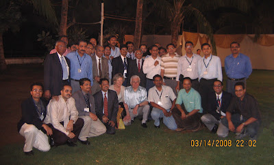
A few snaps at the dinner at Wellington club follows.

From left to right: Bhagwati, Martin, Juha Hernesniemi and umm ... Charlie's angels pretending to be scrub sisters.

Above all, I am sure you would agree with me, that angels are key to any successful surgical mission ;-)

Friday, January 7, 2011

The Plastic Tumor Returns!


Hi!

Any late night suggestions on dealing with this recurrent grade II ependymoma which had been near totally excised and given conformal radiotherapy in 2008 ? You can see the latest scan here,  previous post here and the initial discussion here . Patient presented with headache and has hearing loss on right side on examination. Shunt is functioning and ventricles are small.
Gopalakrishnan.

Friday, November 5, 2010

Haustra Cerebri !

T 2 -weighted axial section of thebrain showing
dilatation of both lateraland third ventricles. b 3D-CISS MRI midsagittal sections of the brain showing aqueductalstenosis with obstructive hydrocephalus.c , d 3D-CISS MRI sagittal sectionsof the brain showingmultiple septaand locules within ventricle appearing as haustrations. The layering fluid within theventricles is probably due to high protein content secondary to infection.

Dhaval Shukla's lateral thought module in action!
Pediatr Neurosurg 2010;46:247–248


Tuesday, October 19, 2010

60th Annual Conference of the Neurological Society of India, Bangalore, 2011

Prof.S.Sampath,
Organizing Secretary, Neurocon 2011.
Prof and Head, Department of Neurosurgery,
NIMHANS, Bangalore.


Click here to register online at www.neurocon2011.com

or Download registration form, fill and mail it the  old fashioned way!


Contact details including email address

Thursday, January 14, 2010

Hyperostosing sphenoid wing meningioma with intra and extracranial parts: Treatment strategy

Please watch the video and post your comments.

Histopath: Transititional meningioma [WHO grade 1]

There was no perception of light in right eye and she was able to count fingers at one feet in left eye . A small residual temporal field of vision exists in left.Papilledema in left fundus has resolved after first surgery. There is optic atrophy in both eyes, severe in right.     


Discussion on this case till now [copied from Yahoo group site activity]

Chief,
Great excision of intracranial portion.
What is the histopathology report?
Its difficult to say how to remove the extracranial part. I feel there could be two ways to excise the extracranial part :
1. endonasal endoscopic excision (navigation assisted)
2. Le-Fort osteotomy and excision.
Let us know further.
Nitin

Hey CHief
Great removal of the intracranial component. Well the extracranial appears to involve the posterior ethmoids more on the left and extending into the sphnoid sinus and uper clivus. I would consider the following options:
1.Transethmoid approach with an ENT collegue, the posterior most may be difficult to reach..
2. ENdoscopic transnasal/transeth moid would also be a good option, best with navigation..
3.If you want an open approach then, extended subfrontal route, you will be able to remove the entire lesion. GOod  ACF base repair is a must
4. Options for involved bone would be to wait for HPE and if grade 1 then wait and follow up oterwise to irradiate, stereotactic radiotherapy. ..
Regards,
MAqsood

Sunday, November 22, 2009

Thalamic lesion for diagnostic biopsy:Dr Nitin's patient


Friends,
This is 35 year old lady with progressive Left third nerve and right hemiparesis over 2 months. She had obstructive hydrocephalus for which she underwent a shunt.
Her CSF did not reveal any malignant cells.
I would like your opinion regarding how to biopsy this lesion. I am strongly considering lymphoma as possibility. She is not immunocompromised and has no other features of systemic involvement.
Unfortunately, our Stereotactic system is not working and patient cannot afford to move out to some other place.
She has a shunt.
I was considering 3 probable options:
1. Block the shunt, repeat scan after 6 hours and if further dilatation of ventricles, do an endoscopic biopsy. Her right frontal horn is still dilated.
2. Subtemporal approach.
3. Posterior transcallosal approach.

Would like your opinions regarding options.

Nitin Garg

Responses

As you suggested , one could do open biopsy of this lesion through several approaches,including frontal transcortical , transcallosal, subtemporal, parietal transcortical, even with modified Poppen's, approach,
I would rather do an endoscopic biopsy through the frontal horn
of right lateral ventricle
I think right lateral ventricle is not adequately decompressed with the VP shunt , which may need to be followed up

I have a suggestion about the discussion here, I think we should avoid showing the identity of the patients here, esp since this is an open forum
Pramod


Dear Nitin,
A very interesting case indeed. It is rather difficult from the images you have uploaded to ascertain the nature of the lesion. is it T2 dark witha spectro pattern favouring a lymphoma. If so you should strongly consider only a minimally invasive biopsy.Stereotaxy is ideal. I am not sure if an endoscopic approach via the right frontal horn will get you to the lesion. You could consider an image guided (ultrasound) biopsy if you have the apparatus.
Having siad that , I have my reservations on the presumptive diagnosis of a lymphoma. The lesion almost looks extra-axial in some of the cuts. More images would be better to determine the exact location to plan the approach. I may be wrong...but its worth a second look...all the best. Keep us posted on the progress.
ali

Some points reg. nitins case.
1.From limited images sent, to me it is an intrinsic lesion though as ali pointed out the left side thalamic- subthalamic- midbrain component has become exophytic in inferior aspect.
2. Fact that it is present in opp. thalamic region makes extrinsic diagnosis fairly less likely.
3. though lymphoma is a good bet based on periventricular intrinsic location and multiplicity another strong contender would be multifocal high grade glial mass-GBM perhaps. The large size of the left sided lesion in my opinioin makes me think of nonlymphomatous possibility.
4.Reg. approach, this is an ideal case for a)Frame based stereotactic surgery or b)Navigation based frameless stereotaxy.
I would be loathe to pursue craniotomy for just a biopsy, however for circumstances u have mentioned u need to do open surgery, I think post. transcallosal would not be my choice.
for subtemporal too it would require too much retraction of dominant temporal lobe and would risk jeoparadising Labbe'.
If u r comfortable with endoscopy go ahead but 6 hours will not suffice to dilate the ventricles and u would not see the lesion if the foramen monroe is not well dilated to pass thru safely.
Best wishes and keep us updated.
Satish
I agree with pramod that we should maintain anonymity of pt. identity.

Monday, November 16, 2009

Brain stem glioma: surgical approach?

14 year old girl with swallowing dysfunction, gaze paresis, facial nerve paresis, and mild quadriparesis.
[Please click the second last button on the you tube bottom bar to get full screen so that you can see in more detail]


Responses

Hi gk.
Good to see you become active again in this site after a hiatus
My thoughts.
1.It is a GBM medulla and pons extensively involved whatever the treatment options excercised, prognosis is grave with few weeks of survival, that being the case, would do everything to stabilise/improve quality of life.
2.would go with retro sigmoid app, need to work deep between cranial nerves, between lower cranial and 7,8th complex and then between 5th and 7th too. Realistically looks like a modest decompression is all that is possible without worsening neuro status.If you could hit the cyst at the summit would be good to reduce mass effect.
3.Temporal horns are getting big, is there significant hydro?
Another development in treating GBMS in general, Avastatin(anti angiogenic) has shown good promise adding few weeks to months for new/recurrent GBMs
Best wishes
Satish Sathya


Dear GK,
I think the retrosigmoid approach is the best to reach and do whatever decompresion that is possible. Probably decompressing the cyst and taking a biopsy is all I would attempt surgically , given the location and malignant nature of the lesion.
regards,
Maqsood

I agree with Dr Satish about its dismal prognosis regardless of the histopathology and extent of surgical resection.The tumor has significant ventrally exophytic component with areas showing some sort of margin from the brain stem.I would attempt to decompress this part of the tumor as well as the necrotic/cystic component through a retrosigmoid/far lateral approach

Pramod

Saturday, November 14, 2009

Olfactory groove meningioma with sinus invasion: Dr Maqsood's patient

Dear all,

This is a 48 yr old lady presenting with long standing anosmia of 8 years and recent adult onset seizures. She has no deficits except bilateral anosmia. Attached is the contrast MRI.
Kindly advise regarding the approach to take out the tumour completely including that of the paranasal sinus part.
I am planning an extended subfrontal approach.
Will an endoscopic endonasal approach be required to mobilise the lower component?
Regards

Dr.Maqsood


Hi Maqsood

The sinus component looks very large and there also appears to be hypertelorism. Is it evident clinically?

Have you considered a transnasal biopsy of the tumor before definitive surgery? It might not be a meningioma at all and even if it is it might be atypical or malignant.

Approach wise -
Extended subfrontal should be able to remove the tumor completely, IMO. Needless to say, reconstruction of the ACF base will have to be meticulous.

All the best with this one. Do let us know how it goes.

Regards

Arvind

Dear Chief, i think the extended subfrontal approach should be enough for the lower part as well, in this case.

Dr. Anirban Deep Banerjee
Neurosurgeon( NIMHANS)

Dear Dr Maqsood,
Thank you for illustrating such a case. To me bi-frontal, or even unilateral sub-frontal approach may be enough for the intracranial part. It is a more or less olfactory groove meningioma invading the base. If you notice, there is no major edema around, denoting the presence of an arachnoid plain which will make your dissection a little easier. The endonasal part may be approached from above, being intra-tumoral all the time during dissection; or may be approached with a combined endo nasal approach. The absence of signal voids intratumorally is a good sign of safe dissection. The most important is the plan of reconstruction, to provide a good base reconstruct and to prevent fistula formation later on. Lastly, a good look at the hormonal profile of the patient is a mandatory pre- and post-operative task.

Looking forward to hear from you.
Thanks
Mohamed Mohi Eldin , MB-BCH , M.Sc., MD
Prof. of Neurosurgery, Faculty of Medicine, Cairo University, Egypt,


Dear maqsood,
Agree that extened transfrontal is ideal for excising tumor completely.
I disagree need for biopsy, regardless of path, and beingn or atypiacal or even nonmeningioma makes no difference for need for complete excision,
Would take a large pericranial graft going behind skin incision and keep duragen plus or eqvivlaent as standby.
you would be surprised at the extent of access from above once intraranial component is removed.
Bewre of retraction, use less or none as bothfrontal lobes are already edematous, give good amount of steroids perioperatively.
Best wishes
Le us know the outcome.
One of our members(not sure if he is in the group) will be salivating seeing this MRI as it is a good and challenging case to do entitely endoscopically by extended transnasal approach(he is our desi amin kassam!).
Satish

Dear Maqsood,

I would agree with Dr Satish that an extended subfrontal approach would suffice in this case. the approach is able to access lesions in the frontoethmoids and nasal cavities. it is only when the leson involves the maxillary sinuses (which is not the case here) that an additional nasal approach (median maxillectomy or an endoscopic approach ) may be needed. This looks like a meningioma (and hence a biopsy is really not required)and I feel the bifrontal approach also provides for an excellent (and a very important) repair of the base.
all the best

Ali

Wednesday, June 24, 2009

How can I become a neurosurgeon? Should I take up General surgery first?

How can I become a neurosurgeon? Should I take up General surgery first?

This are exact same words that an MBBS student asked me the other day. He didnt tell me why he wanted to take up neurosurgery. He said he didn’t have any particular reason. Or maybe he didn’t want to tell me. In any case, it didn’t matter to me. Rational thought and expressed reasoning are not for everyone. For some it’s a calling and that’s fine. That's legit.

More importantly, we have to answer the second question: Should he take up general surgery first? If one has decided on taking neurosurgery as the end speciality, does one really need to go through three years of general surgical training before going through another round of entrance test and three more years of dedicated neurosurgery MCh training?

Lets break this question up…

Why is neurosurgery different from any other surgical field?

I feel that it is because the surgical motor skill that is required is quite different. It is unlike any of the catching-a-bleeder-tying-it-and-dissecting sort of thing that you do in general surgery.
Joints and muscles maketh the man!

May I take the liberty to propose a ‘Motor classification of surgeons” depending on the movements that are required at various joints?

1. If you are a ‘’shoulder-surgeon”, you are a good orthopedician
2. If you are an “elbow-surgeon”, take up general surgery, surgical gastro or cardiothoracic surgery
3. If you are a “wrist-surgeon”, its plastic surgery for you.
4. If you are good with fine finger generated surgical movements, neurosurgery and microvascular surgical fields may be good for you. You are basically restricted to metacarphalangeal and interphalangeal joints and rest of the joints are better stabilized and rested.

Neurosurgery is also not for the claustrophobic and the impatient. Expect narrow corridors and long hours on the operating microscope.

Neurosurgery is not for the morbidity-phobic surgeon either. It’s a fact that despite all the care one takes, one might end up injuring a patient forever… and many patients in a career. This happens in neurosurgery more than any other surgical field, mainly because of the density of functional tissue in the operative field.

Again back to the second question, does one really have to prime oneself with general surgery before taking up neurosurgery?

I believe that it is not necessary. If you have really decided on taking up neurosurgery, why not plunge straight in and save at least one year and be more focused on the subjects that matter – neurology, neuroradiology and operative neuroanatomy.
I am sure that many of you will not agree with me. What about ‘the broader outlook’ to patient management that a post MS general surgery resident is supposed to possess? May be there is a difference. May be you are better off managing a multiply injured patient. But how often has one managed a patient with blunt abdominal trauma and head injury and how often have you operated on a blunt injury patient while managing head injury? May be one can better diagnose the condtion and manage shock and resuscitate better. But does this really require three years of learning hernioraphy, mastectomy and abdominoperineal resection?

But there are caveats. It’s possible that a person who is post MBBS may have deep, nagging doubts throughout the five years whether the decision he has taken was too brave and whether he is up to it. And in the unlikely and unfortunate event of dropping out of the course, one will have nothing but MBBS left even if you have spent many years in the course. Yet, once he has completed the course he might be more focused on the subject and will have saved one year.

Skill, of course depends on the resident.

Lets see another angle to this question.

What type of residents do consultants prefer? For example, Sree chitra [SCTMST] favours post MS candidates of late. I’m not sure of the reasons. May be someone can enlighten on this point.

Friday, January 23, 2009

Is choice an illusion? What next for this nine year old girl?

This nine year old was investigated for 3 months history of defective vision and progressive headache and vomiting. She has primary optic atrophy in right eye [no PL]. She underwent a right ventriculoperitoneal shunt procedure in emergency setting for raised intracranial tension. She does well in school and has intact higher intellectual functions.


The preoperative scans are in the video. Pause the video to make your preoperative diagnoses. What might be the two most likely diagnoses?
Un-pause to see the post operative scans and suggest the next steps. What do you think are the treatment choices?

1)Imaging follow up, and if there is progression consider radiotherapy/Re-excision (which one?)
2)Radiotherapy now and re-excision if progression
3)Chemotherapy?
4)Re-excision now

For those of who with limited bandwidth, get a better connection to see the video! [just kidding... please see the attached jpegs: preop and post op]

Discussion on treatment options for thalamic low grade astrocytoma

What are the treatment options for this 30 year old lady with progressive hemiparesis.
Stereotactic biopsy is suggestive of low grade astrocytoma. Please advise. Am attaching post-GD MRI images.

-Nitin Garg


> From: brmrao1963 To: neurosurgery_ nimhans@. ..:
Sunday, January 18, 2009 11:55:26 AMSubject: [neurosurgery_ nimhans]
Re: Question of the week: Left thalamic low grade astrocytoma
>

Dear Nitin,
The images reveal a multifocallesion which is occupying a spread-out real estate in the ganglionic
and deep grey region abutting the internal capsule. While I do
operate micro surgically on thalamic lesions,the usual indication is
for a single mass. You can use a therapeutic test with dexamethasone
to find out if the weakness is reversible or not. If the deficit is
not reversible with steroids,then it is unlikely that surgery will
do so.In such a scenario, it maybe enough to subject her to
radiation and chemotherapy( some low grade astros do respond to
chemo)taking into consideration QOL issues.If there is a reasonable
chance of reversal of deficit you operate on her(transcallosal Vs
transcortical_ the lesion is spread-out quite laterally and may be
difficult to access transcallosally) . The point to be considered is
that the deficit can be created by a surgical adventure also!If you
have access to DTI please do it to map internal capsule fibers and
their course. If they are involved by the tumor, Surgery will not
have much to offer.

Ravi

To: neurosurgery_ nimhans@. ..: gopalakrishnanms@ ...: Sun, 18 Jan
2009 08:01:34 -0800Subject: Re: [neurosurgery_ nimhans] Re: Question
of the week: Left thalamic low grade astrocytoma

Dear Nitin,
Some questions worth pondering on ...
1. Is the histopath accurate? Does it warrant a review?
2. Is that low grade a sampling effect?
3. Is it possible that this multiple looking entity is finger like fiber tracking [and fiber destroying]
projections of a more sinister glioma or perhaps a different diagnosis considering the edge enhancement and central hypodensity,edema and may be acute (is it?) clinical history?

Regards,
Gopalakrishnan.

In neurosurgery_ nimhansATyahoogro ups.com, Dr.Nitin Garg wrote:
>
>
Thanks for the comments. On replying to both Prof Ravi Mohan Sir's
and GK's queries,

1. The onset was not acute. Patient developed the symptoms over 2-
3 weeks. The weakness is Grade 3-4.
2. During STB, the fluid obtained was xanthochromic. The biopsy
was from wall.
3. There has been no history of fever or other symptoms of TB.
4. Patient has been on steriods for almost 4 weeks. Neurologically
is status quo.
5. Shall try to get DTI.
>
Thanks.
Nitin


Hi Nitin,

I agree with GK , with images you provided(infiltrati ve,multifocal,
heterogenous lesion) , it is worthwhile reviewing the
histopathology, often sampling error from such a large tumor can
occur with stereotactic biopsies,biopsies from the periphery
sometimes show atypical astrocytes and pathologist may label it as
low grade astrocytoma. If this biopsy is from a representative area,
this is being a low grade tumor in a young patient , I would
certainly offer surgical treatment with an aim to maximum
debulking/excision without causing any more neurological deficits
(limitations -if tumor is infiltrative) .With tumor location, I would
intially consider doing a posterior interhemispheric
transventricular approach.But as Dr Ravi Mohan Rao rightly said,
lateral most portion will be difficult to reach with a midline
approach. The residual tumor could be reached through a separate
transcortical approach, aiming for gross total excision, esp if it
is turned out to be a low grade neoplasm.If it is a malignant
glioma , I would be less aggressive in the surgical approach.
Diffusion tensor image could be helpful in locating the white matter
tracts - esp for studying the location of internal capsule,optic
radiation etc.fMRI should be done to locate the speech and motor
areas, esp if you are planning for transcortical approach.
Primary aim of the surgery in this patient should be reducing the
tumor burden, not improving her neurological functions

Pramod

I thank all for the opinion.
Shall get the HPE reviewed and decide about the further mgt later. Shall keep you all informed about the progress.
Bye.
Nitin

[images and clinical details courtesy of Dr Nitin Garg]

Final update on this patient from Dr.Nitin

Friends,
I had posted a case of left thalamic tumor sometime in December. The STB was reported as grade 2 astrocytoma. The patient was planned for RT but was lost to follow up. She now came after 6 months with features of raised ICP. Rpt imaging showed significant frontal extension and midbrain and pontine extension of lesion.
We did a frontoparietal craniotomy, middle frontal gyrus approach and tumor decompression. Intraop impression was high grade glioma. Post-op she recovered with persistent hemiplegia and some aphasia.
The final histopathology has been reported as anaplastic oligodendroglioma. Looking back at the images and some of the comments posted, this looked malignant at that point and may have a sampling error. MRS would have helped (we dont have it in Bhopal at present).
presenting this case for followup.
Bye.
Nitin

Discussion on treatment options for growth hormone secreting residual pituitary macroadenoma

[please see previous post to see the clinical details of this discussion.

From: Arvind Bhateja To: neurosurgery nimhansATyahoogroup
Sent: Monday, December 22, 2008 11:48:42 AM
Subject: Re: Question of the week. Gh secreting residual.
Hi GKand Happy new year everyone!

In my opinion, the best option would be to re-operate this gentleman via an endoscopic trans-sphenoidal which should be able to decompress tumor around the carotid and cav sinuses.
To tackle the bleeding one would probably need to have thrombin solution or floseal.
Its probably the best way although many would find this option more painful.

If endoscopic is an issue an orbitozygomatic with an interdural approach to the cav sinus might also do the job, but it would be overkill for such a small residual.

Almost surely he is going to require hormonal control with cabergoline post-operatively, which might also aid tumor regression if there is still a small residual.

Thanks GK for posing us these teasers and looking forward to some interesting responses!

Arvind

Dr Arvind Bhateja
Consultant Neurosurgeon and Spine Surgeon,
Sita Bhateja Speciality Hospital
Bangalore 560025, INDIA

From: Dr Pramod Pillai To: neurosurgerynimhans
Sent: Tuesday, December 23, 2008 7:04:55 PM
Subject: Re: Question of the week. Gh secreting residual.
Dear All
Merry Xmas and happy new year!!!

I would still try to get his preop and follow up images, see how this tumor behaved over the last 1 year.
If this is just a residual tumor with no significant growth over the last one year, tumor control wont be my concern, instead I would try to treat him
medically with a combination of GH receptor antagonist , pegvisomant with Sandostatin or Somatuline.then follow him with serial images and GH/IGF levels.Patient' s affordability could be an issue

Otherwise , I would rather offer him radiosurgery of his parasellar tumor, it looks like there is enough space between the optic pathway the superior margin of the tumor( we need atleast 2mm distance) , but unlike the nonfunctioning tumors, functioning tumors need higher radiation dose , but there are several methods by which we can reduce dose to the optic pathway to below 8Gy.Nevertheless, there is risk to radiation injury to the cranial nerves in cavernous sinus region, but risk is definitely less than with any surgical approach .Another issue with radiosurgery is latency period , for therapeutic response, during which patient need to be on medical therapyagain. There could be small bit of tumor on the left parasellar region as well?

I wont recommend surgery for this patient , no single approach or surgery is going to cure him off his disease without cranial nerve morbidity, I am also concerned that tumor was encircling the cavernous carotid.

Pramod

From: gopalakrishnan ms
To: neurosurgery_ nimhans
Sent: Tuesday, December 23, 2008 11:53:59 AM
Subject: Re: [neurosurgery_ nimhans] Question of the week. Gh secreting residual.

Please note: The current MRI was done recently [within three months]. Preoperative GH levels were around 40 at the time of primary surgery one year back. An immediate post op MRI was not done. So this is basically a hormonally active residue. He can afford either surgery or radiosurgery (if offered at concessional rate as is done in NIMHANS) but not expensive drugs for an indefinite period.

Shall i take the liberty of adding a sub-question? :
1. What if there is no visible tumor remnant, yet the GH level is high? Is radiosurgical targetting of the entire sella an option?
Wish you Merry Christmas!
GK

From: Dr Pramod Pillai To: neurosurgery_ nimhans@yahoogro

In that case , I would go ahead with radiosurgery, hoping that it provides a hormonal remission so that he can be off the medication later on
The reported remission rate with radiosurgery is about 30-70 %.You do not have to include sella in the target in this case since there is hardly any tumor in the sella.It looks like previous surgeon had tried to preserve the normal pituitary with an intact pituitary stalk( how is his other endocrine functions?)
In rare situations, with all failed therapies with no radiological evidence of tumor in pituitary gland and if you are unable to find another source esp with Cushing's disease, sometimes pituitary gland can be targeted for radisurgery.
If you still like to do surgery on him, carefully study his vascular anatomy.With limited images you provided, I still feel there is encircling of ICA by the tumor , even there is reduced caliber of ICA on the right side. I still feel it is less likely that we can surgically cure him of the disease without causing any additional morbidity.


--- On Thu, 12/25/08,
From: brmrao1963
Subject: [neurosurgery_ nimhans] Re: Question of the week. Gh secreting residual.
To: neurosurgery_ nimhans@yahoogro ups.com
Date: Thursday, December 25, 2008, 6:14 PM
Wishing All of You a very happy and prosperous new year!
I am joining the discussion late. The patient in question has a residual secreting growth
hormone adenoma in the paracavernous area ,probably in the cavernous sinus.
Now the ideal treatment of a GH adenoma residual would be surgery-
1.endoscopic approach
2.miniorbitozygomat ic approach (interdural) .

The drawback of endoscopic approach is this tumor will require an extended endoscopic
approach .This require the surgeon to be extremely conversant with the approach-not
practical for many of us,though I personally do endoscpic pituitary surgery.
This tumor can be removed by an frontotemporal interdural approach which can be
conducted with minimal morbidity. The cranial nerves III and IV can be traced intradurally and interdurally to spare them from injury. I can speak with confidence regarding this approach as I am very familiar with the nuances of this approach.

This tumor cannot be treated with radiotherapy- period!

GKS is an option but may fail. ICA stenosis is a well known complication of GKS. With in adequate contouring cranial nerve palsies are possible!
So the 2 options are interdural approach Vs GKS. My vote is for interdural approach and this will not be overkill as high GH can kill!;patient cannot afford long-term drugs and GKS may or may-not succeed!
Cheers!
Ravi Mohan Rao

Hi all, I agree with Dr. ravi mohan Rao. this can be approached with either extended endoscopic approach or orbitozygomatic. OZ approach is really not morbid procedure as we have done for cavernous sinus tumors. I feel its morbid for the surgeon if u dont have themicro saw drill. not morbid for patient.
GK thanks for posting such cases
regards

Dr.Praveen Ganigi
Consultant Neurosurgeon & Spinal Surgeon
Narayana Institute Of Neurosciences,
Bangalore- 560099

Further update on this patient [Nov '09]
Thank you for all your suggestions...
I had taken a decision to send the patient for gammaknife radiosurgery. [Radiosurgery is not available in jipmer as of now] However, there were some apprehension of cranial nerve deficits, especially optic nerve injury - and the quantum of risks that was explained to the patient was unacceptable to him.
He underwent stereotactic radiotherapy at CMC vellore two months back. He is under follow up. Symptoms persists.
Personally I feel he will eventually require radiosurgery at a future date when the risks of cranial nerve morbidity will be even higher due to previous irradiation. (or microneurosurgery) Ill update you on his response ... though that will be many years away.

Sunday, December 21, 2008

Residual Gh secreting pituitary macroadenoma


What treatment do you suggest for this 30 year old man with a residual Growth hormone secreting pituitary adenoma? He was operated [trans-sphenoidal excision] one year back and his GH level is 28 ng/ml. [i dont have pre-op scans, as he was operated elsewhere]


What do you suggest?

1]Re-surgery [What approach?]
2]Radiosurgery [what are the risks of cranial nerve palsies due to proximity to cavernous sinus and optic nerves, What are the chances of hormonal remission? Target includes sella?]
3]Radiotherapy [Is this an acceptable option considering the time to remission?]

Tuesday, September 16, 2008

Neurosurgery is Carpentry.


I mean, aren't the similarities striking? At least, thats what it looks like from this early colour photograph of a carpenter at work on Douglas Dam, Tennessee in 1942. Look at all that blood, er, dirt on his scrub suit.

How tough can these two professions be? Depends on what your work is...A mayster does rough carpentry, a finish carpenter does exact work, and then there is the miyadaiku - the temple carpenter of Japan who use tools quite distinct from others, like a saw that cuts on the pull and he prefers to do the finest most delicate and aesthetic work in sitting position! And 98.5% of carpenters are men according to wikipedia. [95% of neurosurgeons are too...]
Find out! try your hand at carpentry! Be sure to protect your fingers though.

So, what carpenter are you?

Image source: [original] http://hdl.loc.gov/loc.pnp/fsac.1a35241 [Retouched one is available at wikipedia]

Tuesday, September 9, 2008

Ependymoma: the plastic monster, Outcome

Lesion: WHO Grade II Ependymoma
Residual tumor volume: 1.74 ml
Please watch the video for details ...
ThankYou, Nitin, Ari and Mithun for your thoughts.

Friday, August 22, 2008

Exoskeleton in neurosurgery

Well, I hope you don't think this is too weird.

But how many times have you wished your arms don't get tired while maintaining a particular posture while operating? Hand rests are there, but they don't move with your limbs. You lose time and patience adjusting them.

I was just thinking, wouldn't it be nice if you could have a operating suit (air conditioned one, of course) that adjust its external rigidity to enable one to effortlessly maintain different body postures for prolonged periods?

Is this possible?

Certain animals have muscle tissue that interlock without using further energy . Details escape my memory ... read it in some old physiology book . But hey, we are humans, and we need technology.

I guess we can use some form of electrorheological fluid or elctroactive polymers within the suit that instantaneously harden on applying a voltage. This should keep the total weight of the suit low. Of course we need lots of microprocessors and voice activated controls etc, but then these are easily done. It could even dampen out any unnecessary tremors or potentially hazardous inadvertent movements. {You could program a constrained movement zone at and near the operating field} Well that should definitely help if your assistants tend to fall asleep on your Leyla ;-)

By the way, did you know that chocolate is an electro-rheological fluid? No! no edible suit for you!

This still doesn't completely solve the issue of fatigue because there is no escaping the effort involved in lifting your arms against the pull of mother earth and maintaining it there with all the weight of the suit. [yeah... i know the French have removed a cyst in microgravity environment, but most of our patients are still or terra firma.

What about a powered exoskeleton for the 'supersurgeon'.


This is a Japanese Cyberdyne robosuit HAL. [image courtesy Cyberdyne Inc]

Some of the military guys have all the luck researching such cool stuff.

Just watch this video. [courtesy:berkeleybionics.com]


How much will you pay for a whole body powered exoskeleton neurosurgical operating suit which comes sterile and air conditioned. You don't even have to scrub! Just slip into it and plug in your fuel cells. yes, heads up display, and built in coffee maker are included.

More seriously, a non-obtrusive and slender powered exoskeleton which you can easily strap on to your body could be useful.

Now, where did you say the patent office was? $-)

Saturday, August 16, 2008

How do you deal with this plastic monster?

Lets plan an assault on this tumor!
Please go thru' the MRI pics in the video. The kid is 6 years old with a few months of raised intracranial pressure and nasal twang since two weeks. He has nystagmus but no gross deficits.
What do you suggest?


What would be the surgical approach? positioning?
What about adjuvant therapy - if completely excised/if not/in either case?
And what do you think is this tumor? (ok...I know... it looks obvious)

Sunday, July 20, 2008

How to upload an operative video to neurosurgeryatnimhans blog

This small clip should make it clear.
You need to have admin privileges as an author to add content once you have joined this team blog.

Friday, May 16, 2008

WFNS Education Course and Prof RF Spetzler’s Hands on practical course, May 2008, Goa, India: A review


Great opportunity to listen to some of the renowned experts of the field like A de sousa, Bricolo, Black, Spetzler, Kato and others. It’s a good thing… The hands on cadaveric workshop was little bit of a dampener. Too less time to dissect in a toxic, hot formaldehyde rich atmosphere straight out Venus. That’s probably why the video link was getting cut most of the time?! And why just OZ and far lateral? Eleven thousand rupees is a lot of money if you are not used to earning in tons of Dollars. You deserve a better deal.

By the way, no one came from nimhans. Mais pourquoi? Guys, this is Goa! Missing the workshop is pardonable but not the party. Do get hold of Spetzlers course DVD. The interactive DVD with pictures taken with a Zeiss MKM robotic scope with multilayer Image reconstruction technique is impressive.

Some other desirable stuff/ideas to consider:

Indocyanine green angiography

Spetzler Mallis disposable bipolar leads and lots of ice cubes!

Just edited this movie. Enjoy!


Tuesday, March 18, 2008

M. Gazi Yasargil. Ginde oration and Microneurosurgery CME, Bombay hospital


Amazing energy and intensity at 83!

Some of us could catch up with him at the banquet: Diane and MGY

Shall I quote some of his advices/exhortations?

"Sell your car and buy a CUSA!"
"no nimodipine, no triple H, they just dont help"
"Don't believe in these publications, don't believe in the books, don't believe me either"

His operative video DVD will soon be available. May be, we can watch the op-videos of those scary post fossa AVMS which he didnt have time to show.

A few things to remember...

  • Practice in a cadaver lab... especically microvascular skills
  • Mercilessly coagulate aneurysms in addition to clipping them : this is complete treatment.
  • Have a good set of bipolars, suction tubes and the finest softest cottonoids- don't hesitate to use them in large numbers
  • Have a Leyla retractor but just dont use it. [same goes for assistants]
  • As far as possible, operate on the rich and the famous [Italians] ;-)
A truly memorable event.

Sunday, January 6, 2008

Some more pics ... Neurocon '08


Focal cellular hyperplasia matters... but how do I convince the sheep?... Manish

find the rest at yahoo group site.

At the Taj Mahal ... Neurocon 08